Describe How an Image Formed on the Retina Compares
6Describe the lacrimal system. The process by which this occurs is known as accommodation and will be discussed in more detail in the next part of Lesson 6.
Physics Of The Eye Physics Ii
Now make the actual measurements of image distance and image size and compare them with your predictions.

. This process is called refraction. Although the image formed on the retina is inverted but our brain interprets this correctly Ie. Like camera the eye lens forms a real and inverted image of the object on retinathe optic nerve carries it in the form of signals to the brain.
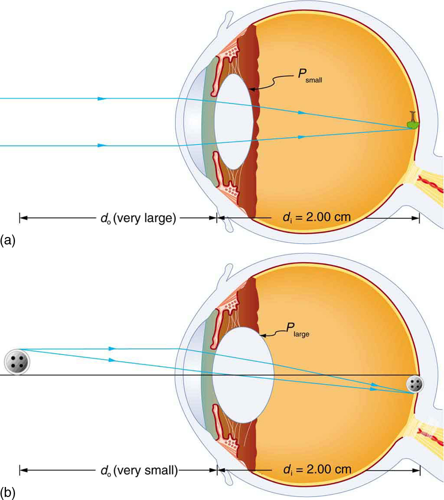
The two eyes look at images from slightly different. Measure the height of your object and predict the size of the image which would be formed with the object placed as described above. A case 1 image is formed when d o f and f is positive as in Figure 10a.
Use this activity to see how retinal image size depends upon the distance and the size of the object. Describe how an image formed on the retina compares with its actual appearance in the outside world. A summary of the three cases or types of image formation appears at the end of this section A different type of image is formed when an object such as a persons face is held close to a convex lens.
Small muscles attached to the eye lens are constantly changing the curvature of the lens. Describe abnormal findings of tissue color that are possible on the conjunctiva and sclera and describe their significance. Cameras and eyes contain convex lenses.
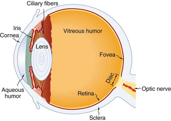
The fovea is a region of the retina that is specialized for high visual acuity and color perception. From the center of the optic nerve radiates the major blood vessels of the retina. When the ball ball leaves your hand its retinal image is a certain size defined by the angle the size of the obect has at your eye.
Note that we use the law of reflection to. We cannot get such image using a concave lens because the concave lens always produces the diminished image. The cornea and lens of an eye act together to form a real image on the light-sensing retina which has its densest concentration of receptors in the fovea and a blind spot over the optic nerve.
The iris acts like the diaphragm of camera. The image formed is left-right reversed and inverted. Bending the light rays is necessary in order to focus an external objects image and emit the light just behind the lens at an area called the focal point.
They convert light into biological signals. Fortunately the image is a real image - formed by the actual convergence of light rays at a point in space. An image is formed in the human eye when light passes through the pupil is refracted by the lens and is absorbed by pigment cells in the retina altering the pigments and triggering neurons to fire.
The right way up. The figure shows the virtual image formed by the convex lens and real image formed by a. What kind of image is formed.
The figure below presents the formation of an object image into a. The particular arrangement of the stimulated cells is interpreted by the brain into a separate image for each eye. To understand how this happens consider Figure.
Lens depends on the lens used and the distance from the object to the lens. When an ophthalmologist uses an ophthalmoscope to look into your eye he sees the following view of the retina Fig. Rods and cones are structurally compartmentalised.
An object in the upper temporal visual field of the right eye reflects its image onto the lower nasal area of the retina. Yes an enlarged image is formed. In humans rods cone and retinal ganglion cells are the 3 main photoreceptors.
The retina is the thin light-sensitive membrane lining the inner eyeball- towards the back of the eye. The Formation of Images on the Retina Because light rays diverge in all directions from their source the set of rays from each point in space that reach the pupil must be focused. Because the image need to be in the same place on the retina this focal distance becomes smaller for closer objects.
They are very sensitive They can be triggered by a single photon also. As the ball moves away from you the retinal image size of the ball shrinks as the distance increases. The image is then focused on the retina.
The retinal output fibers leave at a point in the retina called the blindspot. Also the image looks smaller than the external object in view. When light goes into your eye it.
The formation of focused images on the photo receptors of. Answer 1 of 2. A camera or human eye.
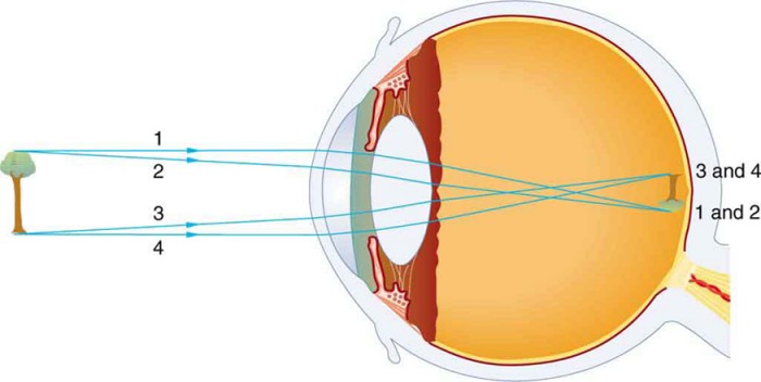
Figure 21 contains an overview of. For this arrangement the image distance and linear magnification of the image are given by. The image formed on the retina is upside down and reversed from its actual appearance in the outside world.
We shall refer to these as case 1 images. What we see through our eyes are inverted image but the image formed on the retina will be in the two dimensional form It is our brain which helps us to interpret the 2D images as 3D images. Answer 1 of 4.
This allow us to see the world as a sharply focused image. Describe how an image formed on the retina compares with its actual appearance in the outside world. The opening at the center of iris is.
Images in a plane mirror are the same size as the object are located behind the mirror and are oriented in the same direction as the object ie upright. Describe how an image formed on the retina compares with its actual appearance in the outside world The image formed on the retina is upside down and reversed from its actual appearance in the outside worlds ie. The power of the lens of an eye is adjustable to provide an image on the retina for varying object distances.
Two rays emerge from point P strike the mirror and reflect into the observers eye. Images through lenses as real virtual erect or magnified. The image formed on the retina is upside down and reversed from its actual appearance in the outside world.
In the center of the retina is the optic nerve a circular to oval white area measuring about 2 x 15 mm across. Rods are narrow and are present throughout the retina. The bundle of output fibers is called the optic nerve.
Quite conveniently the cornea-lens system produces an image of an object on the retinal surface. Photoreceptors in the retina are classified into two groups named after their physical morphologies. Provides constant irrigation to keep the conjunctiva and cornea moist and lubricated.
So this is a convex lens. Rod cells are highly sensitive to light and function in night-vision whereas cone cells are capable of detecting a wide spectrum of light photons and are responsible for colour vision. The type of image formed by a convex.
2D images will be converted into electrical signals and these signals will be. Photoreceptors are responsible for visual phototransduction.
Physics Of The Eye Physics Ii
Physics Of The Eye Physics Ii
Image Formation In The Eye And The Camera
Image Formation In The Human Eye A When An Object Is Observed At A Download Scientific Diagram
No comments for "Describe How an Image Formed on the Retina Compares"
Post a Comment